espresso sun
How Berkeley and Biohub built a laser phase plate the size of an espresso cup, and opened a window into 90% of human biology cryo-EM could not see.
June 11, 2026
A laser 100 million times brighter than the Sun bounces between two mirrors ten thousand times and is focused to a spot thinner than a human hair, and the whole apparatus is less than four inches wide (Biohub). It fits inside a microscope like a cartridge, which is to say the intensity of the Sun contained in something about the size of an espresso cup.
On June 11, researchers at UC Berkeley and CZ Biohub announced they had built a device the field had spent fifteen years calling impossible, a laser phase plate that lets scientists see proteins they have never been able to see before.
More than 90% of the proteins inside a human cell are too small for cryo-electron microscopy to image clearly (Biohub press release), and every one of those proteins is a potential disease mechanism and a potential drug target, so the laser phase plate is the first credible path to making the majority of them visible. Three papers dropped the same day, one in Science, one in Nature Communications, and one preprint on bioRxiv, and two microscopes in the world now have this technology installed.
The ceiling
Cryo-EM won a Nobel in 2017 and has since become the backbone of structural biology. You flash-freeze a biological sample in thin ice, fire an electron beam through it, and reconstruct the 3D structure from thousands of images, and the technique has driven drug design, revealed the architecture of huge protein complexes, and become standard in pharma.
But cryo-EM has a hard limit: biological samples are nearly transparent to electrons, so contrast is extremely low for small proteins, and the field compensates by averaging thousands of images of identical copies of a molecule. That works when the molecule is large and rigid, but when it is small, below about 100 kilodaltons, there is not enough signal to align the images and the reconstruction falls apart.
The result is that cryo-EM can resolve about 10% of the human proteome in purified form and, inside intact cells, below 1% (Biohub blog), meaning the remaining 90-plus percent has been invisible.
Physicists identified this problem in the 1940s, when Fritz Zernike showed you could solve it in light microscopy by inserting an optical element that converts phase differences into brightness differences, work that won him the Nobel Prize in 1953. Scientists immediately wanted to do the same in electron microscopy, but they could not, because every material placed in an electron beam gets bombarded, charges, and degrades, and the closest anyone got was the Volta phase plate in the 2010s, a thin carbon film that worked intermittently but was unstable and blurred high-resolution detail (Petrov et al., Science, 2026), leaving the contrast problem open for eighty years.
Holger
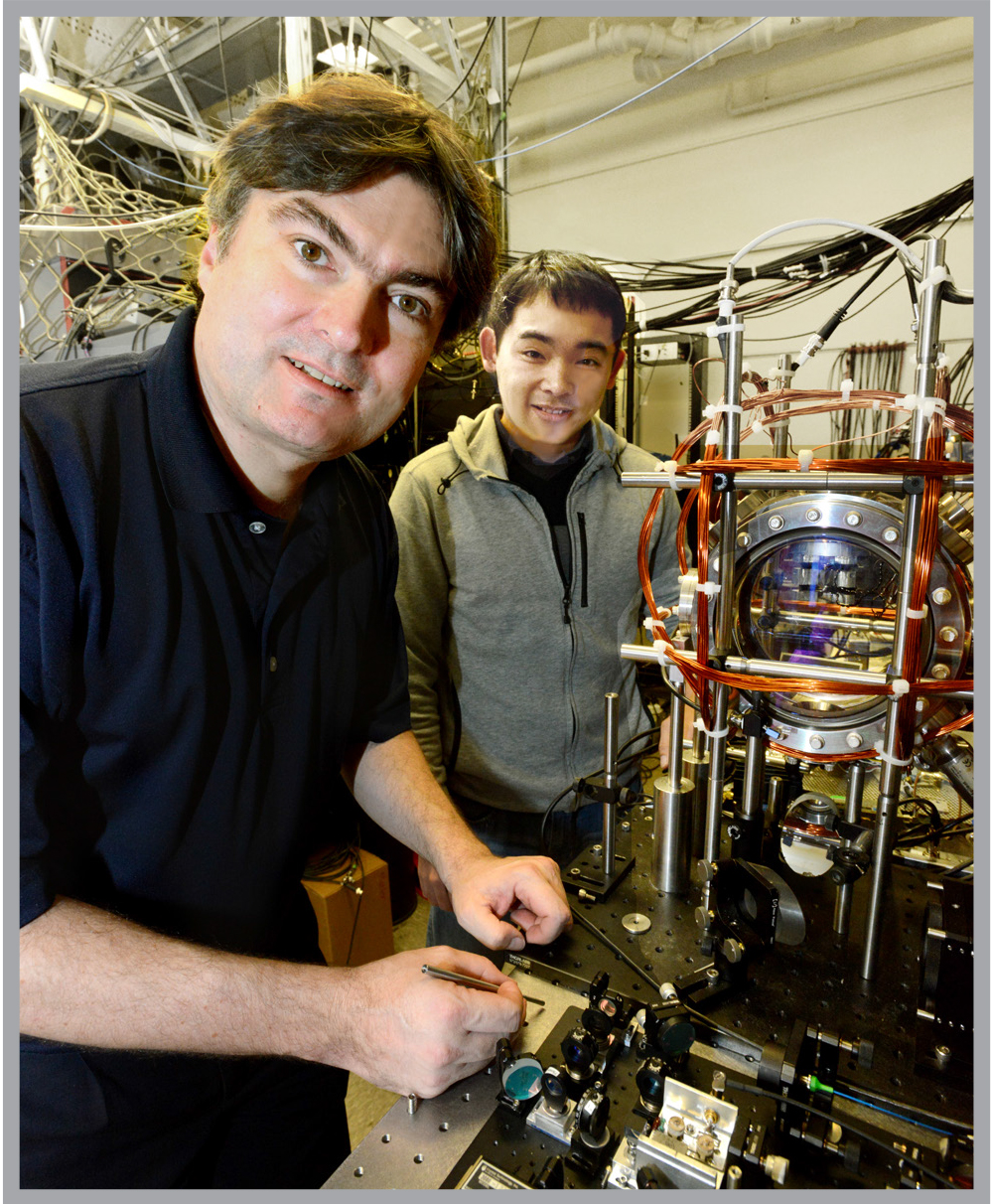
Holger Müller is a professor of physics at Berkeley, a Packard Fellow, and the kind of person whose lab page reads as three research groups stapled together. His primary work is atom interferometry, building quantum instruments that measure gravity and probe the Standard Model. He filed his first patent at fourteen, grew up in Germany, studied at Konstanz and Humboldt-Berlin, joined Steven Chu's group at Stanford, and arrived at Berkeley in 2008 (UC Berkeley Physics).
The laser phase plate came out of a collaboration with Robert Glaeser, who matters here because Glaeser is one of the founders of cryo-EM itself. Now 88 and professor emeritus at Berkeley, he spent 56 years at Lawrence Berkeley National Lab, and in the 1960s he established that radiation damage was the limiting factor for cryo-EM resolution and showed that freezing specimens reduced it. The 2017 Nobel Committee credited his work (LBNL).
In 2010, Müller and Glaeser published a paper proposing a fix (New Journal of Physics, 2010): instead of inserting a material phase plate into the electron beam, insert a focused laser beam. Light interacts with electrons through the ponderomotive force, and if the laser is intense enough and stable enough, you can shift the electron wave's phase without putting any solid material in the beam path, which avoids contamination, charging, and degradation.
The catch is that light barely interacts with electrons, so the laser would need to be extraordinarily intense, focused to a micron-scale spot, held stable for hours, and positioned to within nanometers of the electron beam. Müller's design is a Fabry-Perot cavity, two concave mirrors bouncing a laser back and forth roughly 10,000 times and amplifying the intensity each pass until the focused light reaches about 350-400 gigawatts per square centimeter (Biohub press release), which is one hundred million times the intensity of the Sun's surface concentrated into a spot one-thousandth the width of a human hair.
Atomically smooth
The engineering tolerances are what earned the "impossible" label. The mirrors must be polished to a surface roughness below one angstrom, roughly the diameter of a single atom, and aligned to within a thousandth of a degree, because a single speck of dust landing on one will absorb the laser, burn, and destroy the mirror. The laser beam and electron beam must be co-aligned to within 50 nanometers on a 500-nanometer standing wave and held there for half-hour stretches of data collection (Biohub press release).
One peer reviewer on the Biohub preprint reportedly objected that nobody would ever be able to build it (Labcritics).
Müller spent over a decade building a working prototype, and in 2021 Biohub made a major investment through a CZI-funded grant to purchase a Thermo Scientific Krios, customize it for the laser phase plate, and push the prototype into a modern instrument (Biohub blog). Müller calls this microscope "Theia," a Formula 1 microscope with extra electron optics that outperform a standard Krios before the laser is switched on (Labcritics).
The entire laser apparatus fits in a module less than four inches wide, or as Biohub puts it, "about the size of an espresso cup" (Biohub blog), and from the outside the microscope looks like any other Krios.

Two days of silence
The Berkeley team ran paired experiments, laser off and then laser on, on the same sample and the same microscope. Postdocs Jessie Zhang and Petar Petrov imaged aldolase, a standard benchmark, and hemoglobin, the oxygen-carrying protein in blood, which at 64 kilodaltons sits right at the lower edge of what current cryo-EM can handle.
Technician Jonathan Remis took the data and ran it through the reconstruction pipeline over the weekend, then went silent while Zhang and Petrov waited two days. When Remis came back, he said, "What if it's so good I want to make sure it's correct before sharing it with you?" (Biohub blog). With the laser on, resolution improved by up to 44% (UC Berkeley News), and features that had been blurry snapped into clear structure. The improvement was largest for the hardest sample, hemoglobin, and smallest for the easier one, aldolase, which is exactly what the theory predicted (Petrov et al., Science, 2026).
Meanwhile, Biohub built a second, different design called the crossed laser phase plate (xLPP), with two laser beams in an X-shaped configuration, each in its own cavity. The dual-beam approach splits power between two beams, which reduces mirror stress and catastrophic-failure risk, and suppresses ghost images, faint duplicates that can swamp biological signal. They imaged apoferritin at 1.8 angstroms, approaching the theoretical resolution limit of the technology (Yu et al., bioRxiv, 2026).

The workshop question
In the winter of 2019, 25 physicists and engineers gathered in San Francisco for a workshop on the future of electron microscopy (Biohub blog). They had hit a barrier: despite better cameras, better software, and better sample prep, cryo-EM still could not produce high-contrast images of proteins inside a cell.
Stephani Otte, Biohub's VP of imaging science, organized the meeting and has described her operating principle as, "If somebody says it can't be done, that's the biggest motivating factor for me" (Biohub blog). Toward the end of the workshop, she asked the group which single tool, of all the unmet needs in the field, would move science forward fastest.
The answer was nearly unanimous: Müller's laser phase plate, though most of them added that they did not think it could actually be built. Biohub funded it anyway, and over the next seven years they backed Müller's work at Berkeley, built on it, and developed the dual-laser xLPP at their own lab in Redwood City, and both microscopes have now produced results.
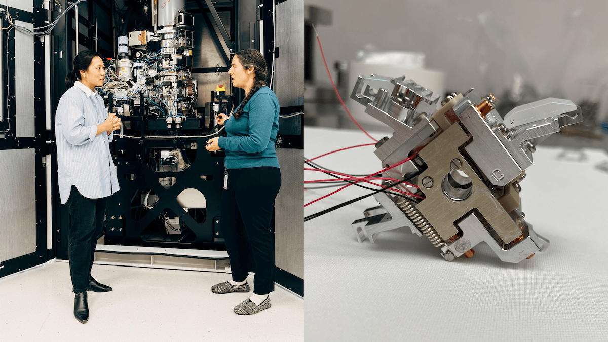
The key people on the Biohub side include David Agard (founding scientific director of imaging), Bridget Carragher (founding technical director and co-lead of the Dynamic Structural Cell Biology group), Pavel Olshin (the engineer who built the xLPP hardware), and Otte herself. Carragher has described the current state as, "It's like seeing first light through a telescope. The science it enables, that comes next" (Biohub blog).
Virtual cells
In April 2026, Biohub announced the Virtual Biology Initiative, a five-year, $500 million effort to generate the cellular datasets needed to build predictive AI models of the human cell, with $400 million for internal data generation and technology and $100 million to seed a global data effort, aiming at "virtual cells," AI systems trained to simulate how cells work, malfunction in disease, and respond to treatment (Biohub announcement).
The laser phase plate fits into the Virtual Biology Initiative as a data-generation instrument: if cryo-electron tomography with the LPP can image proteins inside cells at near-atomic resolution, those images become training data for AI models that predict protein interactions, map disease mechanisms, and accelerate drug design, which means the laser phase plate is not just a better microscope but an attempt to make the interior of the cell machine-readable.
Biohub is releasing its cryo-ET data openly through the CryoET Data Portal, which already hosts tens of thousands of annotated tomograms, and open data is the part of this that scales a field rather than just a lab.
First light
The next frontier is cryo-electron tomography, imaging proteins not in isolation but inside intact cells in 3D and in context. Agard calls this "where we'll see the really huge wins for cell biology" (Biohub press release), Carragher says they expect to be collecting cryo-ET data by the end of 2026 (Biohub blog), and Alexis Rohou, director of cryo-EM at Genentech, told Science magazine that "any technological gain that makes your resolution better is always welcome" (Science).
It took fifteen years to get here, with three papers landing in a single day, mirrors polished smoother than anything in nature, and what may be the brightest continuous-wave laser ever built, backed by a philanthropy willing to spend hundreds of millions on the bet that if you can see what is happening inside a cell, you can change it. Jeremy Axelrod, a PhD student in Müller's group, graduated in 2024 with a thesis titled "A Laser Phase Plate for Transmission Electron Microscopy" (Müller Group), and Jonathan Remis went quiet for two days because the results were better than anyone had prepared for.